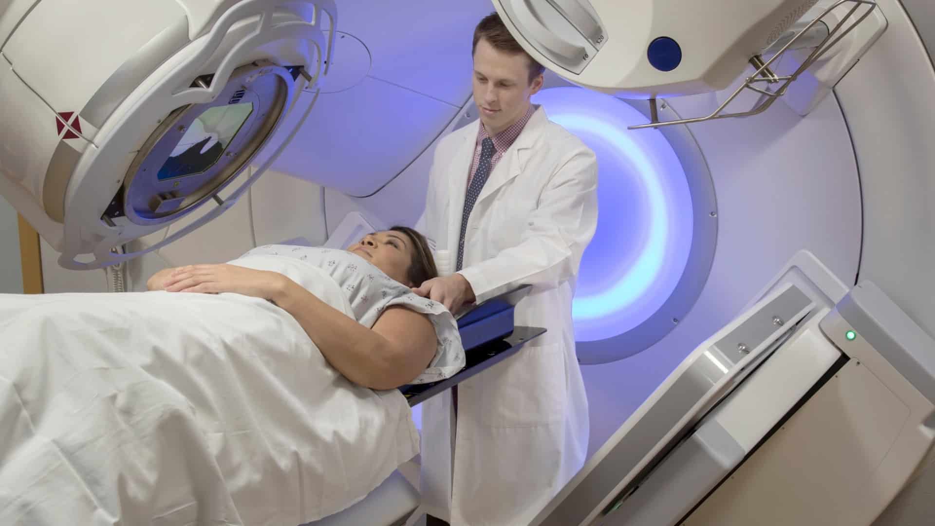
How Radiation Treatment Has Evolved to Protect Healthy Tissue?
When you think about radiation therapy, you might picture powerful beams hitting both the tumor and everything around it but that’s no longer the full story. Today’s treatments use detailed scans, computer-guided planning, and tightly shaped beams to focus radiation where it’s needed and spare healthy tissue as much as possible. Still, not all techniques are the same, and understanding what’s changed can influence the choices you make next.
How Modern Radiation Therapy Protects Healthy Tissue
Modern radiation therapy is designed to damage cancer cells while limiting exposure to nearby healthy tissue. Before treatment begins, CT or MRI scans are used to define the exact size, shape, and location of the tumor and to identify surrounding organs. This information allows planners to shape the radiation dose so that critical structures receive as little exposure as possible.
During treatment, techniques such as intensity‑modulated radiation therapy (IMRT) and image‑guided radiation therapy (IGRT) deliver beams from multiple directions to concentrate dose in the tumor and reduce dose to healthy tissue. Stereotactic approaches provide even more precise targeting for selected tumors by using highly focused beams and strict patient positioning.
Treatment plans also specify the total dose, the size of each fraction, and the margins around the tumor, taking into account established dose limits for different organs. In some cases, hypofractionated schedules are used to complete treatment in fewer sessions while remaining within safe dose constraints.
Follow‑up imaging is then used to assess the tumor’s response and to guide any necessary adjustments in future treatment.
Where a tumor sits close to the heart or spinal cord, some patients are also assessed for particle treatment rather than X‑ray beams. Clinical oncologist Dr James Wilson offers proton beam therapy in London for lung and other thoracic cancers, delivered through Proton International London at UCLH; because protons stop at the tumor instead of passing through it, the dose reaching the heart can be reduced by as much as half. His treatment page sets out which patients are usually considered for it and how the course is scheduled.
From Early Radiation Beams to 3D Tumor Targeting
In the early 1900s, radiation therapy relied on broad, relatively imprecise beams derived from early X‑ray machines and radium sources. These treatments primarily targeted rapidly dividing cells, such as cancer cells, but the technology didn't allow accurate shaping or confinement of the dose.
As a result, radiation often extended beyond the tumor, exposing surrounding healthy tissue to significant doses and increasing the risk of skin burns, tissue damage, and long‑term side effects.
How Imaging Guides Safer Radiation Treatments
As imaging technology has advanced, it has turned radiation therapy from a relatively imprecise approach into a more accurately guided process that better limits exposure to healthy tissue.
CT and MRI scans allow the care team to see the tumor and surrounding organs in fine detail when planning each course of treatment.
During image-guided radiotherapy (IGRT), images are taken in the treatment room to verify the tumor’s position and the patient’s alignment.
The setup can then be adjusted by a few millimeters if needed.
This method, first developed for use in the brain in the late 1960s, is now applied to many areas of the body to direct radiation beams from multiple angles, align them with the planned target, and reduce the dose to normal tissues as much as possible.
Planning Radiation Doses to Protect Nearby Organs
When your care team plans radiation treatment, they do more than target the tumor. They also calculate how much radiation nearby normal organs can safely receive. Using imaging studies such as CT and, when appropriate, MRI, they outline the tumor and identify “organs at risk.” Each of these structures is assigned a dose limit based on known tissue sensitivity and evidence linking dose levels to specific side effects.
Radiation is typically delivered in multiple smaller treatments, called fractions. This fractionation allows healthy tissues some time to repair between treatments, while cancer cells accumulate damage more readily. When treatment requires higher doses per fraction over a shorter overall course, planners usually apply stricter dose limits to nearby organs to reduce the risk of exceeding safe exposure levels.
IMRT, IGRT, and SBRT: Precision Radiation in Action
With current technology, radiation therapy can be directed very precisely to the tumor. In intensity‑modulated radiation therapy (IMRT), the radiation beam is divided into many small beamlets whose intensity can be adjusted from multiple angles. This allows higher doses to be delivered to the tumor while reducing the dose to nearby normal organs.
Image‑guided radiation therapy (IGRT) incorporates frequent imaging, such as X‑rays or CT scans, before or during treatment sessions. These images help confirm the tumor’s position and allow the care team to account for day‑to‑day variations in patient setup or internal organ motion.
Stereotactic body radiation therapy (SBRT) is used in selected cases to deliver relatively high doses of radiation in a small number of treatments. It relies on accurate imaging, careful patient positioning, and motion management techniques.
Proton and Particle Therapy to Spare Healthy Tissue
Beyond shaping and directing X‑ray beams with techniques such as IMRT, IGRT, and SBRT, radiation oncology can also use different types of particles to reduce radiation exposure to healthy tissue. Proton therapy uses positively charged particles that deposit most of their energy at a specific depth in the body, known as the Bragg peak.
Because the dose falls off quickly beyond this point, proton beams can reduce radiation to structures located behind the tumor, such as the spinal cord or certain critical organs.
Other particles, such as carbon ions and other heavy ions, deliver denser ionization along their paths. This may increase biological effectiveness in some tumor types, particularly those that are less responsive to conventional X‑ray radiation, although access is limited and evidence continues to develop.
In all cases, accurate imaging, treatment planning, and daily patient positioning are required to align the beams with the target and maintain the intended balance between tumor control and normal‑tissue protection.
How Today’s Radiation Lowers and Manages Side Effects
Although radiation still works by damaging cancer cells’ DNA, modern techniques are designed to better protect healthy tissue than older approaches.
Treatment planning typically uses CT or MRI scans, and image guidance helps align the radiation beams with the tumor’s exact shape and daily position.
Methods such as intensity-modulated radiation therapy (IMRT) and stereotactic body radiation therapy (SBRT) allow the dose to be concentrated more precisely in the tumor, reducing exposure to nearby organs.
Many treatment plans now use hypofractionation, which delivers a higher dose per session over a shorter overall treatment period, when this has been shown to be safe and effective for a given cancer type.
Clinicians monitor the total radiation dose a patient receives over time and consider prior treatments when planning new courses of therapy.
They also work to prevent and manage side effects by using measures such as specialized skin care, medications to control nausea, and targeted support for bowel, mouth, swallowing, or breathing problems, depending on the area being treated.
Using Precision Radiation to Treat Metastases Safely
In treating cancer that has spread to other parts of the body (metastatic disease), modern precision radiation techniques aim to control tumors while limiting exposure to nearby healthy tissue. Common approaches include intensity-modulated radiation therapy (IMRT), image-guided radiation therapy (IGRT), and stereotactic body radiation therapy (SBRT). These methods use multiple beam angles and advanced planning software so that the radiation dose closely conforms to the shape of each tumor.
SBRT delivers highly focused, relatively high doses of radiation over a small number of treatment sessions (fractions). This approach can provide effective local control of selected metastases while reducing the volume of normal tissue receiving high-dose radiation. To improve accuracy, the treatment team typically uses CT and/or MRI imaging, along with small skin marks or internal markers, to ensure the body is positioned consistently for each session.
Some treatment plans use hypofractionation, in which each dose is higher and the total number of sessions is lower than in conventional radiation schedules. Ongoing clinical trials are evaluating how best to use these techniques for different types of metastatic cancer, with the goals of improving tumor control, minimizing side effects, and protecting nearby organs.
Questions to Ask Your Care Team About Safer Radiation
A clear set of questions can help you understand how your care team plans to treat the cancer while limiting exposure to healthy tissue. You may want to ask how the treatment plan is designed to reduce radiation dose to normal tissues and how imaging is used to guide accurate targeting while minimizing dose to nearby organs.
Consider asking about the specific technologies being used, such as intensity-modulated radiation therapy (IMRT), image-guided radiation therapy (IGRT), or stereotactic body radiation therapy (SBRT), and how these approaches improve precision compared with older, less targeted methods.
It can be helpful to clarify how your team sets dose limits for sensitive structures (for example, the spinal cord, lungs, heart, bowel, or salivary glands, depending on the site) and what side effects are most likely for your particular cancer location and treatment field.
You may also wish to ask how potential side effects will be monitored and managed during and after treatment, including what symptoms you should report right away.
Request information about the schedule for follow-up visits and imaging, how your response to treatment will be assessed, and the typical time frame in which changes in the tumor (such as shrinkage or stability) are usually seen for your type of cancer.
Conclusion
You’ve seen how far radiation therapy’s come in protecting your healthy tissue while still powerfully treating cancer. With better imaging, smarter planning, and precise delivery, your team can focus radiation where it’s needed and avoid where it’s not. You’re not stuck with old‑fashioned, “scattershot” treatments anymore. As you discuss options, ask about techniques like IMRT, IGRT, SBRT, or proton therapy so you can choose the safest, most effective plan for you.
TecCast ®
TecCast is a Semi - Dry product which uses a combination of traditional materials and technologically innovative admixtures to produce stone which is without equal.
TecLite ®
TecLite uses a fibre-reinforced mix to produce light weight products which look and behave exactly like traditional cast stone.